Author: Dr. Daphne Chiu, vet at Lynbrook Veterinary
When your dog suddenly skips a step, briefly lifts a back leg, or seems to “pop” back into stride, it may look quirky or harmless. However, these behaviours can be subtle signs of a common orthopaedic knee condition known as medial patellar luxation (MPL).
While this condition may appear minor at first, untreated MPL can lead to:
- long-term pain,
- reduced mobility,
- and the development of arthritis.
Early diagnosis by a vet and appropriate intervention can make a significant difference to your dog’s comfort, mobility, and overall quality of life.
Here’s our guide on MPL: exploring the anatomy of the canine knee, how MPL is graded, the signs to watch for at home, available treatment options, and the potential complications if the condition is left untreated. All written by our experienced Vet, Dr Daphne Chiu.
What is Medial Patellar Luxation?
Medial patellar luxation occurs when the patella (kneecap) moves out of its normal position within the trochlear groove, a shallow V-shaped channel located at the end of the femur (thigh bone).
Normally, the patella glides smoothly up and down this groove as the knee bends and straightens. In dogs with MPL, the kneecap most commonly slips toward the inside (medial aspect) of the leg.
How Common is MPL?
MPL is the most common orthopaedic condition diagnosed in small-breed dogs, such as Pomeranians, Chihuahuas, Toy Poodles, Yorkshire Terriers, and Cavalier King Charles Spaniels.
The condition is often present from a young age due to inherited differences in limb alignment, bone shape, and muscle pull.
An experienced veterinarian can assess your young dog and advise if this condition is present and likely to lead to MPL as they age.
Although MPL is most frequently diagnosed in small dogs, dogs of all sizes and activity levels can be affected, including medium and large breeds. The severity of the condition varies widely, ranging from occasional, mild skipping to persistent lameness, pain, and progressive joint degeneration.
The Anatomy of the Canine Knee (Stifle Joint)
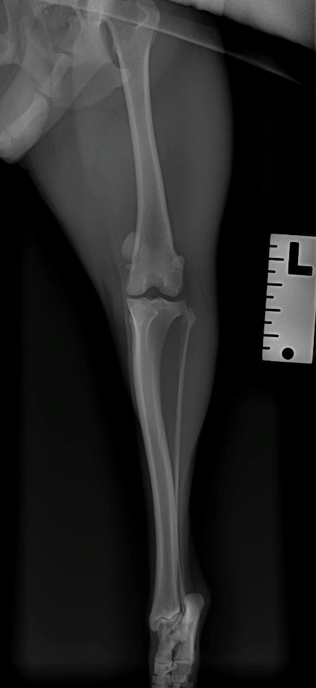
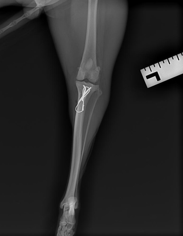
A dog’s knee, also known as the stifle joint, is a complex hinge joint that plays a critical role in weight-bearing and movement. It is made up of three primary bones. The femur (thigh bone), the tibia (shin bone) and the patella (kneecap).
This image would need to be downloaded and supplied by LV. (I cannot find something suitable from Envato or Canva) We can highlight the specific bones that the vet is referring to.
The patella sits within the trochlear groove at the end of the femur and is embedded within the quadriceps tendon. As the quadriceps (thigh) muscle contracts, it pulls the patella smoothly along the groove, allowing efficient knee extension during walking, running, and jumping.
Several structures work together to stabilise the knee, including ligaments (such as the cranial cruciate ligament), joint capsule, cartilage, and surrounding soft tissues. Healthy cartilage lining the joint surfaces allows smooth, pain-free movement and absorbs shock during activity.
In dogs with MPL, this finely balanced system is disrupted. The trochlear groove may be too shallow, the tibial tuberosity may be misaligned, or the quadriceps muscle pull may be directed at an abnormal angle. As a result, when the quadriceps contract, the patella is pulled out of the groove instead of staying centred, leading to intermittent or permanent luxation.
Over time, this abnormal movement causes inflammation, cartilage wear, joint instability, and pain.
The Signs of Medial Patellar Luxation
In the early stages, many dogs compensate remarkably well. Clinical signs vary depending on the severity and frequency of patellar luxation, as well as the dog’s age, size, and activity level.
Common signs owners may notice include:
- Intermittent skipping or hopping on a hind leg
- Briefly holding a back leg up while walking or running
- Sudden extension of the hind limb, followed by normal movement
- Bunny-hopping gait when running
- Reluctance to jump, climb stairs, or exercise
- Stiffness after rest or exercise
- Persistent lameness in more severe cases
Some dogs show signs only occasionally, while others develop continuous lameness and discomfort. Because MPL progresses over time, early signs should not be dismissed as “normal” or age-related.
Grading the Severity of MPL
To guide diagnosis and treatment decisions, our veterinarians (and others) classify MPL using a grading system from Grade 1 (mild) to Grade 4 (severe).
Grade 1
The patella can be manually displaced during examination, but returns to its normal position once pressure is released. Dogs with Grade 1 MPL typically show no obvious signs of lameness and may remain asymptomatic for years.
Grade 2
The patella intermittently luxates out of the groove but may spontaneously return to its normal position. Dogs often show occasional skipping, brief lameness, or sudden extension of the hind limb. Clinical signs are usually intermittent but may worsen with activity.
Grade 3
The patella is permanently luxated but can be manually repositioned into the groove. However, once pressure is released, it quickly luxates again. Dogs with Grade 3 MPL typically show consistent lameness, altered gait, and muscle loss in the affected limb.
Grade 4
The patella is permanently luxated and cannot be manually replaced. Dogs with Grade 4 MPL have significant limb deformities, persistent lameness, and severe gait abnormalities. This grade is often associated with marked bone and soft tissue changes.
Treatment Options: Conservative Versus Surgical Management
Treatment of MPL depends on the grade of luxation and how much the dog is affected.
Conservative Management (Grade 1 to Mild Grade 2)
For mild cases of MPL where lameness is infrequent and there are minimal arthritic changes on x-rays, non-surgical management may be appropriate.
Conservative treatment may include:
- Controlled exercise and activity modification
- Physiotherapy to strengthen supporting muscles Can you link to Jada’s physiotherapy page maybe?
- Weight management to reduce joint load
- Non-steroidal anti-inflammatory drugs (NSAIDs) for pain relief
- Joint-protective supplements to support cartilage health
The goal of conservative management is to minimise inflammation, maintain muscle strength, and slow the progression of joint degeneration. While this approach does not correct the underlying anatomical problem, many dogs remain comfortable for extended periods with appropriate monitoring.
Surgical Management (Grade 2 to Grade 4)
In moderate to severe cases of MPL that are causing pain and discomfort despite conservative management, surgical correction is recommended.
Here’s a helpful short video of what the surgery entails.
Standard Patellar Luxation Surgaery
The primary goal of surgery is to restore normal knee alignment so the patella remains within the trochlear groove during movement. This helps reduce pain, improve function, and prevent further joint damage.
Common surgical techniques include:
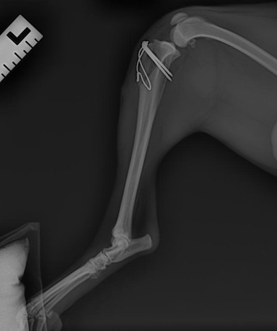
- Trochleoplasty/Sulcoplasty: Deepening the trochlear groove so the patella can sit and glide correctly
- Tibial tuberosity transposition (TTT): Repositioning the attachment of the quadriceps tendon to realign the pull of the muscle
In dogs with more severe deformities of the femur or tibia, additional corrective procedures may be required to address underlying bone alignment issues.
With appropriate surgical planning and post-operative care, most dogs experience significant improvement in comfort and mobility.
Secondary Complications of Untreated MPL
If MPL is left untreated, particularly in moderate-to-severe cases, progressive joint damage can occur over time.
Common long-term complications include:
- Chronic pain and inflammation
- Progressive osteoarthritis
- Persistent or worsening lameness
- Muscle wasting and limb deformities
- Reduced activity and quality of life
Dogs with MPL are also at an increased risk of cranial cruciate ligament (CrCL) rupture. Abnormal limb alignment and rotational forces place ongoing strain on the ligament, making rupture more likely.
Additionally, repeated luxation of the patella causes abnormal friction within the joint, leading to cartilage damage and bone changes. This results in the release of inflammatory enzymes that further degrade joint structures, accelerating arthritis and increasing the risk of ligament injury.
Conclusion
Medial patellar luxation is a common orthopaedic condition, particularly in small-breed dogs, but it is highly manageable with early diagnosis and appropriate treatment. Recognising the signs of MPL and understanding its severity can help prevent progression to painful arthritis and secondary joint complications.
If your dog is:
- skipping,
- intermittently holding up a hind leg,
- or showing any signs of lameness,
we recommend scheduling a thorough orthopaedic examination to assess their stifles and determine the most suitable treatment plan customised for you and your pets’ needs.
Our clinic has extensive experience in diagnosing and treating orthopaedic conditions such as MPL, and we are dedicated to helping your dog regain comfort, mobility, and long-term joint health. Get in touch with our team here, or book an appointment today.